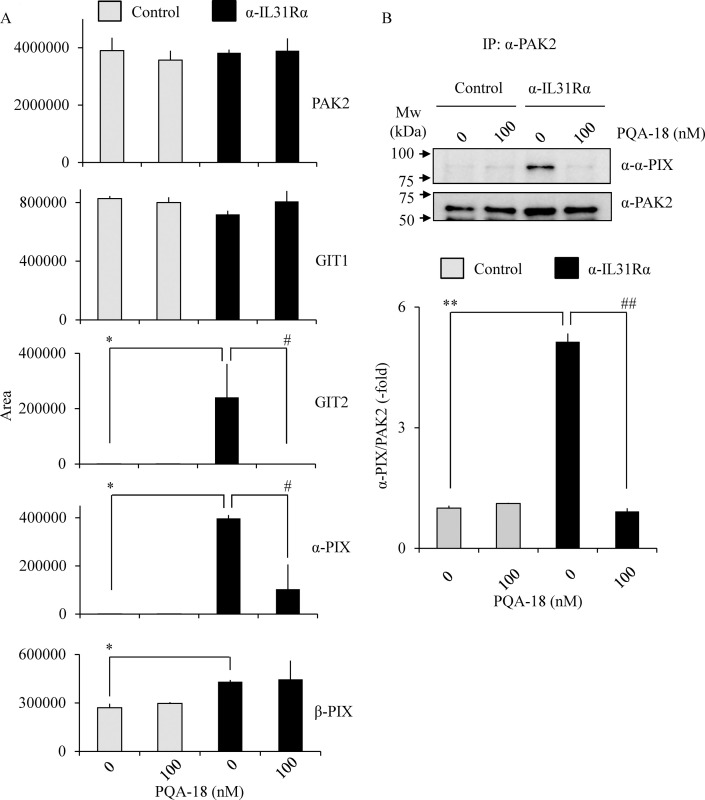
Fig 8. Prevention of formation of PAK2 activation complex by PQA-18.
PAK2 interacting proteins as identified by LC/MS/MS (A). LC/MS/MS analysis was performed using tryptic peptides of purified PAK2 prepared from Neuro2A cells treated without or with PQA-18 in the absence or presence of anti- IL-31Rα antibody using anti-PAK2 antibody conjugated beads. The quantitative data of the peak area of the indicated proteins are shown. Immunoprecipitates were obtained from Neuro2A cells treated without or with PQA-18 in the absence or presence of anti- IL-31Rα antibody using anti-PAK2 antibody conjugated beads, and immunoblotted with anti-α-PIX antibody or anti-PAK2 antibody (B). The representative images are shown (upper), and the quantitative data of the ratios of α-PIX versus PAK2 are shown (lower). Data are pooled from three independent experiments and shown as mean and SD. **p < 0.01, *p < 0.05 as compared with control; ##p < 0.01, #p < 0.05 as compared with anti-IL-31Rα antibody-treated group (one-way ANOVA/Tukey-Kramer post-hoc comparisons).

