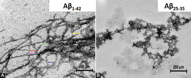
FIGURE 4.

Morphological appearance of amyloid beta (Aβ) fibrils under transmission electron microscope. Aβ fibril aggregates formed from Aβ1-42 peptide demonstrating a “striated ribbon” morphology, with a diameter range from 6 to 15 nm (A), while, Aβ25-35 peptide formed tiny and short Aβ aggregates (B). Scale bar is 200 nm.
