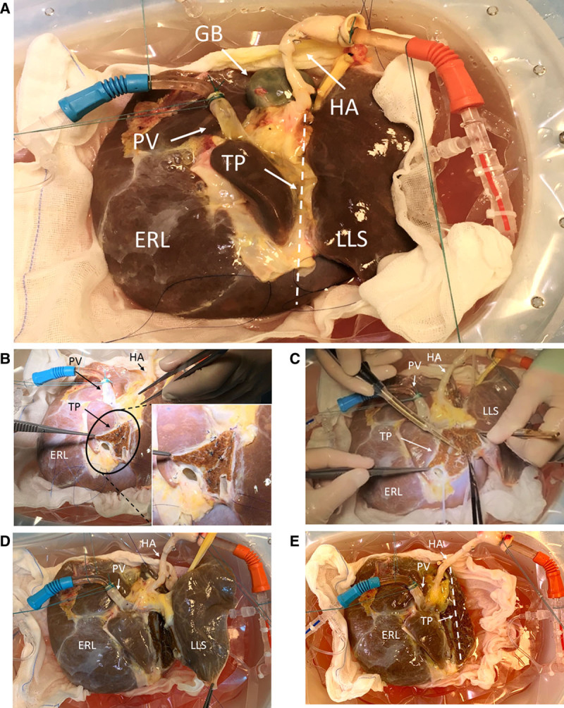
FIGURE 2.

The progression of the split procedure is observable from (A) start of dual hypothermic oxygenated machine perfusion (DHOPE), (B) start of left lateral segment (LLS)/extended right lobe (ERL) liver split with division of the middle and left hepatic vein with magnification of the transection plane, (C) midway through parenchymal liver split using the CUSA device, (D) demonstrating full parenchymal separation of the LLS from the ERL, and (E) showing dual perfusion of the ERL only, after the LLS has been fully removed. CUSA, Cavitron ultrasonic surgical aspirator; GB, gall bladder; HA, hepatic artery; PV, portal vein; TP, transection plane.
