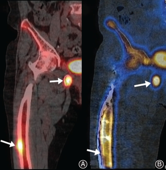
Fig. 7.

Bone single photon emission CT of patient 2. (A) Preoperative nuclear medical images showed hot uptake in the mid‐shaft of the right femur shaft and around the symphysis pubis (arrowhead). (B) Sixteen months after surgery, uptake in the mid‐shaft of the right femur was decreased, but hot uptake was still observed around the symphysis pubis (arrowhead).
