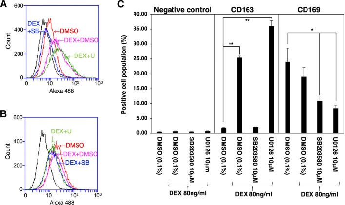
Figure 5.
Flow cytometric analyses of DEX-treated IPKM. IPKM were treated with or without 80 ng/ml DEX for 1 d in the presence or absence of 10 μM SB203580 or 10 μM U0126. DMSO (0.1%) was used as a vehicle control because SB203580 and U0126 were dissolved in DMSO. Then, the cells were reacted with mouse monoclonal anti-CD163 (A) or anti-CD169 (B) antibodies, before being labeled with Alexa Fluor 488-conjugated anti-mouse IgG antibodies (Alexa 488). The positive region was set up to contain about 0.5% of the negative control cells, which reacted with Alexa Fluor 488-conjugated anti-mouse IgG antibodies alone (A and B: black line). Three independent experiments were performed, and the data regarding the percentages of cells in the positive region are expressed as mean±SEM values (*p < 0.05, **p < 0.01 vs. DMSO-treated control) (C). The frequency of CD163-positive cells was significantly higher among the DEX-treated IPKM (A: purple line, and C). The administration of SB203580 completely blocked the effects of DEX (A: blue line, and C). Conversely, the administration of U0126 facilitated the DEX-induced increase in the frequency of CD163-positive cells (A: green line, and C). The frequency of CD169-positive cells was slightly lower among the DEX-treated IPKM (B: purple line, and C). Co-treatment with DEX and SB203580 (B: blue line) or U0126 (B: green line) significantly decreased the frequency of CD169-positive cells (C).

