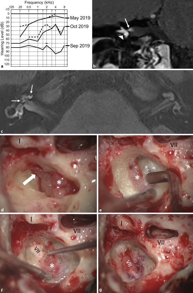
Fig. 5.
Transmacular ISL (patient 8 in Table 2). a Pure-tone audiogram showing fluctuating hearing loss (full line: air conduction, dashed line: bone conduction) most likely based on a secondary cochlear hydrops because of the tumor in the vestibule (c). b MRI (coronal, T1-w + Gd) showing the tumor in the right vestibule (triangle), the ampullary ends of the superior and lateral semicircular canals and extension along the superior vestibular nerve into the IAC (→). c MRI with hydrops sequence (3D inversion recovery, 6 h after systemic Gd application) showing a mild cochlear hydrops (→). d Intraoperative view with the tumor (asterisk) in the vestibule and along the superior vestibular nerve towards the fundus of the IAC (→). e–g The tumor (asterisk) is gradually separated from the facial nerve under facial nerve monitoring control. VII facial nerve, Gd gadolinium, I incus body, w weighted

