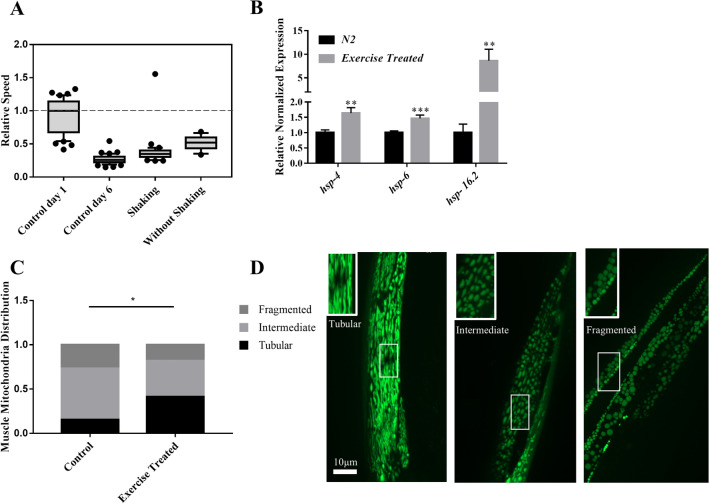
Figure 7.
Effect of Exercise on electrotaxis reponse. (A) Worms were subjected to swimming exercise. A significant increase in the speed was observed using two different treatments (with shaking and without shaking: p < 0.001) compared to same age control. The speed was slower than day-1 controls (p < 0.0001). (B) RT-qPCR analysis of hsp-4, hsp-6 and hsp-16.2 in wild-type adults following 7 days of shaking exercise treatment. The expression of all three chaperones was significantly increased (p < 0.01). (C, D) Muscle mitochondria morphology in day-6 myo-3::GFP(mit) adults after shaking exercise. Animals were placed into three different categories, i.e., having mostly tubular, fragmented, and intermediate (i.e., combination of tubular and fragmented) shapes of mitochondria. These shapes correspond to mitochondrial network being normal (tubular) and defective (fragmented and intermediate). Exercise resulted in significantly more animals having tubular mitochondria (p = 0.045). The numbers of animals were: (A) Control day-1: n = 41, Control day-6: n = 48, shaking: n = 36 , without shaking: n = 14. (B) N2: n = 3 batches, exercise treated: n = 3 batches, (C) Control: n = 23, exercise treated: n = 23. Data was analyzed using one-way ANOVA with Dunnett’s post hoc test (A), one-way ANOVA with Tukey’s post-hoc test (B), and Chi-square test (C).

