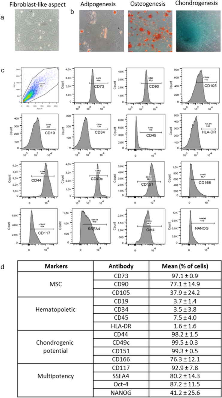
Figure 1.
Characterization of human AFSCs: (a) cells showing adherent growth, exhibiting fibroblast-like aspect (400 ×); (b) differentiation potential in mesenchymal lineage after culture in specific medium (400 ×): adipogenic, oil Red O staining indicating lipidic vesicles; osteogenic, alizarin red staining indicating calcium matrix formation and chondrogenic, Alcian blue staining evidencing glycosaminoglycan presence; (c) immunophenotype analysis of AFSCs by flow cytometry; (d) percentage of cells showing specific markers of MSC, hematopoietic, multipotency, and chondrogenic potential in different samples.

