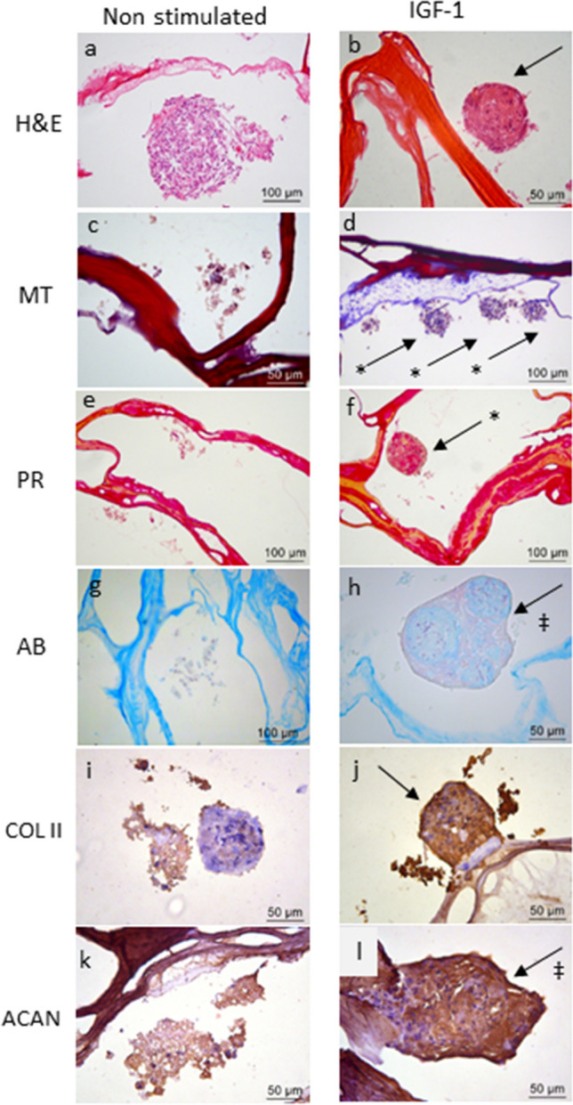
Figure 4.

Chondrogenic differentiation of CD117+ AFSCs seeded into CX scaffold for 21 days with and without IGF-1 stimulation. In the group stimulated by IGF-1 (b,d,f,h,j,l) more compact agglomeration of the cells is observed,with a higher amount of extracellular material compared to that noticed in the non-stimulated control group (a,c,e,g,i,k). H&E staining (a,b); collagen (indicated with *) stained in blue by Masson´s trichrome (MT) (c,d); collagen fibers shown in red by Picrosirius red (PR) (e,f); glycosaminoglycans (‡) stained light blue using Alcian blue (AB) (g,h). Labeling with anti-collagen type II antibody (COL II) (i,j) and anti-aggrecan antibody (ACAN) (k,l) in the stimulated group compared to the control group.
