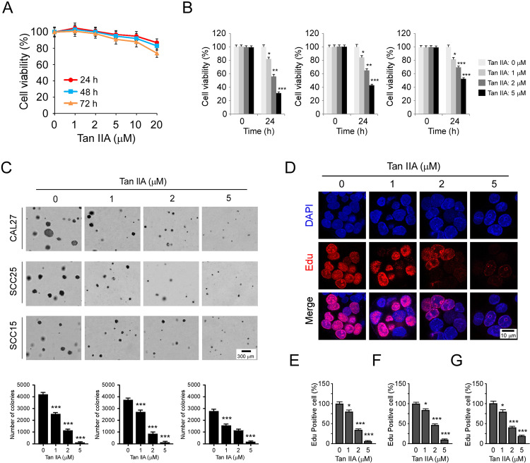
Fig. 3. Tan IIA suppresses OSCC cells.
A MTS assay analyzes the cell viability of hTERT-OME cells with Tan IIA treatment for various time points. B MTS assay analyzes the cell viability of CAL27 (left), SCC25 (middle), and SCC15 (right) cells with Tan IIA treatment for 24 h; n = 3 independent biological replications, one-way ANOVA. C Soft agar assay examines the colony formation of CAL27 (top), SCC25 (middle), and SCC15 (bottom) cells treated with Tan IIA; n = 3 independent biological replications, one-way ANOVA; *p < 0.05, **p < 0.01, ***p < 0.001. D–G Edu incorporation assay and quantitative analysis of the effect of Tan IIA on OSCC proliferation. (D) The representative image of Edu incorporation assay for CAL27 cells. (E–G) Quantitative analysis of Edu incorporated cells for CAL27 (E), SCC25 (F), and SCC15 (G); n = 3 independent biological replications, one-way ANOVA; *p < 0.05, **p < 0.01, ***p < 0.001.

