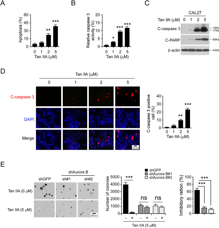
Fig. 5. Tan IIA promotes apoptosis in OSCC cells.
A Flow cytometry examination of apoptotic CAL27 cells with Tan IIA treatment for 72 h; n = 3 independent biological replications, one-way ANOVA; **p < 0.01, ***p < 0.001. B and C CAL27 cells were treated with Tan IIA for 72 h, cell lysate was subjected to cleaved-caspase 3 activity analysis (B) and IB analysis (C). For B, n = 3 independent biological replications, one-way ANOVA; *p < 0.05, ***p < 0.001. D CAL27 cells were treated with Tan IIA for 72 h and subjected to IF analysis with cleaved-caspase 3 antibody. Scale bar, 10 μm; n = 3 independent biological replications, one-way ANOVA; **p < 0.01, ***p < 0.001. E Knockdown of Aurora B decreased the sensitivity to Tan IIA. Soft agar assay analysis of the anti-tumor effect of Tan IIA on shGFP and shAurora B-expressing CAL27 cells (left and middle), the inhibitory ratio of Tan IIA on colony formation was calculated (right); n = 3 independent biological replications, two-way ANOVA, ***p < 0.001; ns, not statistically significant.

