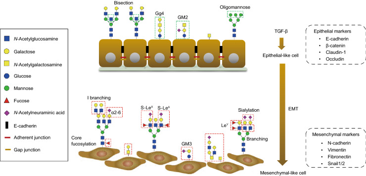
Figure 4.
Glycosylation changes in TGF-β-induced EMT. During TGF-β-induced epithelial–mesenchymal transition (EMT), the epithelial cells lose their cell-cell contact and apical-basal polarity, acquiring a mesenchymal phenotype with enhanced motility and invasion ability. Upon EMT, epithelial markers including E-cadherin, β-catenin, claudin-1 and occludin are downregulated and mesenchymal markers such as N-cadherin, Vimentin, Fibronectin and Snail1/2 are increased. Glycosylation changes occur during EMT. Different types of changes are shown in the red-dashed boxes, highlighting changes in O-glycans and increased expression of branched, core fucosylated and sialylated N-glycans. In addition, the Lewis antigens (S-LeX, S-LeA and LeY) of N-glycans also upregulated within this process. The composition of the GSLs changed, as showed by the depletion of Gg4 or GM2 and expression of GM3 during TGF-β-induced EMT.

