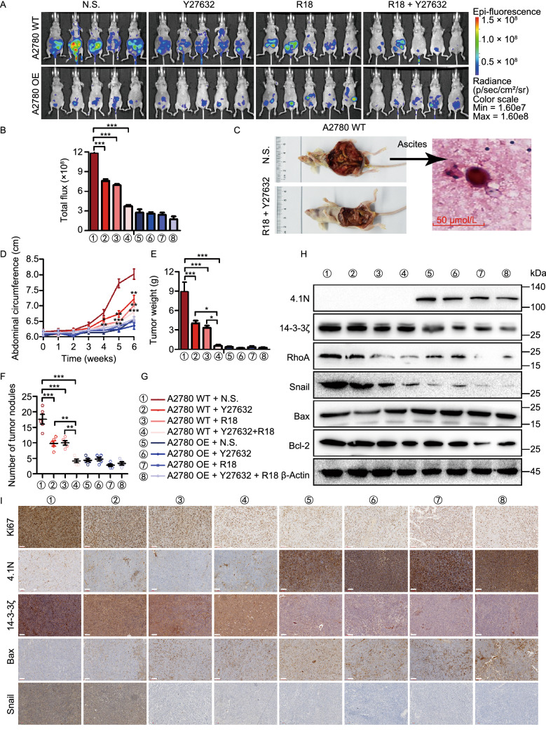
Figure 6.
Loss of 4.1N induces EOC aggressiveness via 14-3-3 expression and entosis in vivo. Peritoneal dissemination nude mouse models were established using A2780 WT and A2780 OE cells treatment with Y27632 and (or) R18. (A and B) In vivo bioluminescent images and quantification of 6 weeks after abdominal injection indicated cells (n = 5, two-sided Student’s t-test). (C) H&E stained sections of abdominal ascites containing examples of entosis structures, as indicated by arrows. Scale bars = 50 μm. Abdominal circumference curves (D), the weight (E) and number (F) of dissected metastatic tumor nodules were quantified. (G) Different colors and number indicate different groups. (H) Protein expression of 4.1N, 14-3-3ζ, RhoA, Snail, Bax and Bcl-2 in nude mouse tissue detected by Western blot. β-Actin was used as an endogenous control. (I) Protein expression of Ki67, 4.1N, 14-3-3ζ, Bax and Snail in nude mouse tissue detected by IHC. Scale bar, 100 µm. *P < 0.05, **P < 0.01 and ***P < 0.001 (Student’s t-test)

