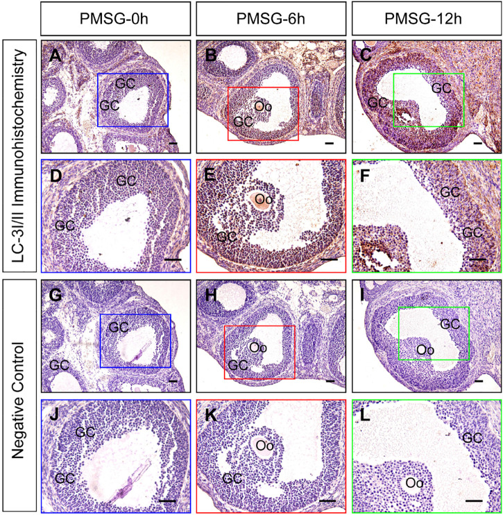
FIGURE 1.
Immunohistochemical staining of LC-3I/II during follicular development. Three-week old female Sprague–Dawley rats were treated with PMSG to induce follicular development. LC-3I/II expression was examined using immunohistochemistry at 0, 6, and 12 h after PMSG treatment. In negative control, slides were incubated with serum instead of LC-3I/II antibody. GC, granulosa cell, Oo, oocyte, Scale bar = 100 μm.

