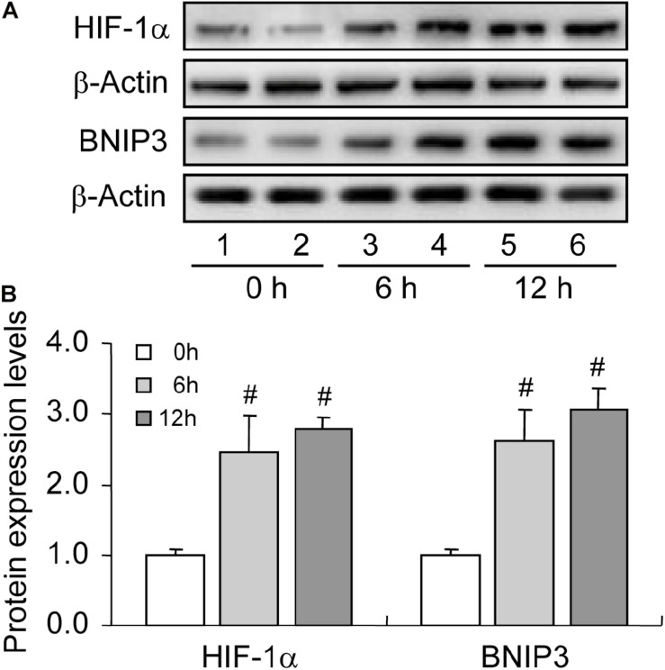
FIGURE 3.
Expression changes of HIF-1α and BNIP3 during follicular development. HIF-1α and BNIP3 expression was examined using western blotting at 0, 6, and 12 h after PMSG treatment. (A) Representative immunoblotting of HIF-1α and BNIP3. (B) Densitometric quantification of HIF-1α and BNIP3. Each value represents the mean ± SE. One-way analysis of variance (ANOVA) was used to analyze the data, followed by a Tukey’s multiple range test. N = 6. #, P < 0.05, vs. 0 h.

