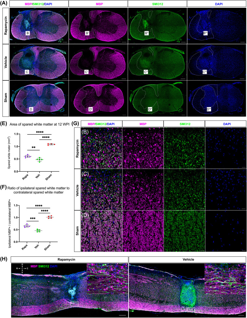
FIGURE 4.
The area of spared white matter after injury. Demyelination was mainly located on the ipsilateral side at the injury epicenter [A, red: myelin basic protein (MBP), labeled myelin sheathes; green: SMI312, labeled axons; blue: DAPI, labeled nucleus]. High magnifications of demyelination are shown in (B–D,G). The area of spared white matter was significantly decreased in the two injured groups, and the area of spared white matter in the rapamycin-treated group was significantly greater than that in the vehicle-treated group (E,F). Longitudinal sections are shown in (H). WPI, weeks post-injury; R, rostral; C, caudal; D, dorsal; V, ventral. ∗∗P < 0.01, ∗∗∗P < 0.001, ****P < 0.0001. Scale bar = 200 μm (A–H) and 20 μm (B–D). Error bars are mean ± SEM.

