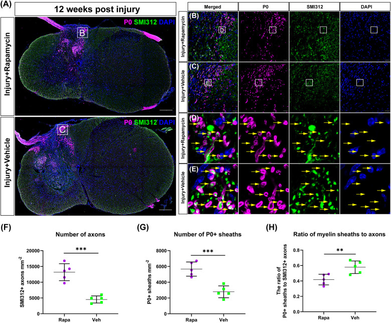
FIGURE 5.
The Schwann cell-mediated remyelination at the injury epicenter of the two injured groups at 12 weeks post-injury. Immunofluorescent staining was used to detect the myelin formed by Schwann cells, and the P0 + myelin was observed around the lesion site and at the nerve roots (A). High-magnification images of (A) are shown in (B,C). (D,E) The representative images of the P0 + myelin (yellow arrows). Quantitation of the SMI312+ axons, P0 + myelin, and the ratio of myelin sheaths to axons were shown in (F–H), and SMI312 + axons and P0 + myelin sheaths were significantly increased in the rapamycin-treated mice when compared with the vehicle-treated mice. The ratio of P0 + myelin to SMI312 + axons in the rapamycin-treated mice was significantly less than that in the vehicle-treated mice. **P < 0.01, ***P < 0.001. Scale bar = 200 μm (A) and 20 μm (B,C). Error bars are mean ± SEM.

