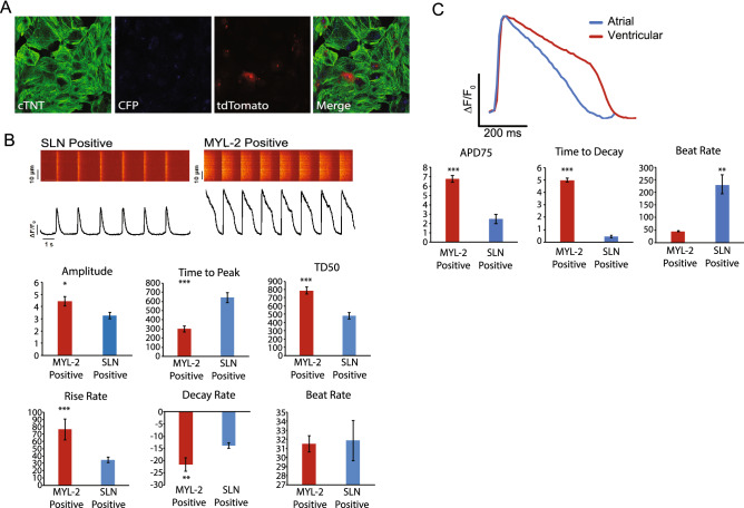
Figure 5.
Characterization of the atrial/ventricular human iPSC Double Reporter Line. (A) Representative immunofluorescence images of day 30 cardiomyocytes from the double reporter system, MYL-2/tdTomato (red) and SLN/eCFP (cyan), immunostained for cTNT (green). (B) Representative calcium tracing and quantification of amplitude, time to peak, TD50, rise rate, decay rate, and beat rate of MYL-2/tdTomato (+) and SLN/eCFP(+) cells (day 30) from the double reporter system. (C) Representative action potential tracing of MYL-2/tdTomato (+) and SLN/eCFP(+) cells (day 30) from the double reporter system separately using voltage activated fluorescent dye and quantification of APD75, decay rate, and beat rate of MYL-2/tdTomato (+) and SLN/eCFP(+) cells (day 30) from the double reporter system.

