Figure 4.
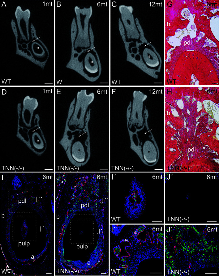
Analysis of lower incisor cross-sections (A–F) μCT scans at the heights of the first molar showed that compared to wildtype (A–C) the incisor dentin layer is much thicker in 1 month old tenascin-W/TNN deficient mice (D, asterisk), in 6 and 12 month old mice the incisor pulp is completely obliterated (E, F, asterisk). A second phenotypic change in the mutant is the wider incisor periodontal space, indicated by an arrow (D–F). However, the periodontal space of the molars seems normal. (G, H) Sirius red stainings showed a significant alveolar bone loss and a replacement by a dense connective tissue in the mutant compared to wildtype. (I, J) An analysis of blood vessels (CD31 in red, smooth muscle actin (SMA) in white, Tuj1 in green) and nerve fibers revealed that in TNN-deficient mice, the remaining pulp tissue does not containing any projections (I´, J´); in contrast the bone replacement tissue contains a well developed network of nerve fibers (I´´,J´´) (a ameloblasts; pdl periodontal ligament; mice, n = 6, scale bar μCT: 0.5 mm, histology: 100 μm).

