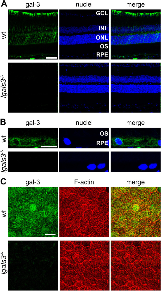
Figure 1.

Gal-3 localizes to the neural retina and RPE in mice. (A) Immunofluorescence staining of gal-3 of retinal sections from wt and lgals3−/− mice shows localization to the inner retina (and they are based on the appearance most likely to be mainly Müller cells), RPE, and choroid. Images are representative maximal projections. Green: gal-3; and blue: nuclei counterstain. GCL, ganglion cell layer; INL, inner nuclear layer; ONL, outer nuclear layer; OS, photoreceptor outer segment layer. Scale bar = 50 µm. (B) Magnified fields of stained sections as in A focusing on the RPE. Scale bar = 10 µm. (C) Immunofluorescence staining of RPE whole mount preparations of wt and lgals3−/− mice show gal-3 in the RPE. Images are representative maximal projections comprising the entire RPE. Green: gal-3; red: F-actin; blue: nuclei counterstain. Scale bar = 25 µm.
