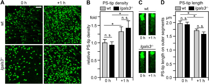
Figure 2.
Diurnal PS exposure at photoreceptor outer segment tips is indistinguishable between wt and lgals3−/− mice. (A) Representative PS-biosensor live imaging of freshly dissected neural retina whole mount of wt and lgals3−/− mice collected within 5 minutes of light onset (0 hours) and 1 hour later (+ 1 hour). Images are maximal projections. Scale bar = 10 µm. (B) Quantification of the frequency of outer segments exposing PS in wt and lgals3−/− mice of samples as in A. Values are normalized to the frequency of outer segments exposing PS in wt mice at light onset. (C) Length of the tip of outer segments exposing PS in wt and lgals3−/− mice at time points as in A. Images are representative maximal projections of PS-biosensor labeling of outer segments in whole mount preparations imaged at high magnification. Scale bar = 2 µm. (D) Quantification of the length of PS-exposing tips of outer segments from images as in C. Bars in B and C show mean ± s. e. m., n = 4; 2-way ANOVA with Tukey's post hoc test; *p < 0.05; n. s., not significant.

