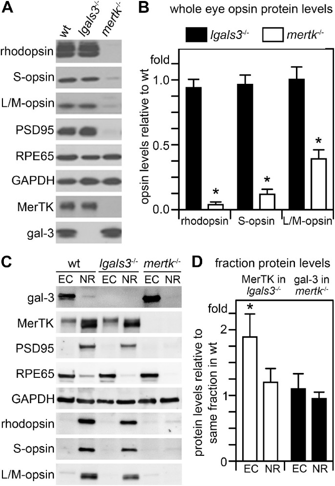
Figure 5.

Unlike degenerating mertk−/− retina, lgals3−/− retina has wt levels of RPE and photoreceptor marker proteins, but gal-3 is elevated in posterior eyecups of mertk−/− mice with retinal degeneration. (A) Representative comparative immunoblots of detergent lysates representing equal fractions of whole eyes without lens with intact retinas of 3-month-old wt, lgals3−/−, and mertk−/− mice probed for photoreceptor and RPE marker proteins and housekeeping proteins GAPDH as loading control as indicated. Probes for gal-3 and MerTK confirmed mouse genotypes. (B) Bars show quantification of average rhodopsin and cone opsin content in lgals3−/− (black bars) and mertk−/− eyes (white bars) normalized to content in wt eyes as indicated. (C) Representative immunoblots of posterior eyecups (ECs) and neural retina (NR) eye fractions probed for proteins as indicated. Probes for RPE65, PSD95, and opsins confirmed fraction enrichment with minor contamination of adjacent tissue as expected of manual dissections. GAPDH indicated protein load. (D) Bars show quantification of gal-3 in mertk−/− eye fractions (white bars) and of MerTK in lgals3−/− eye fractions (black bars), respectively. Bars in B and D show averages ± s. e. m., n = 4; Student's t-test; *p < 0.05. There was no difference in levels of opsins, MerTK or any other marker tested between lgals3−/− and wt tissue. As expected, mertk−/− eyes with ongoing retinal degeneration contained dramatically reduced levels of opsins and other neural retina markers but normal levels of RPE marker RPE65 and elevated levels of gal-3.
