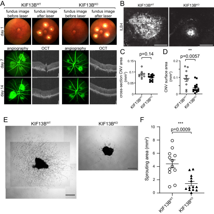
Figure 4.
Genetically modified model of KIF13B knockout shows inhibition on laser-induced CNV and sprouting from choroidal vessels. (A, B, C, D) Impaired CNV in global KIF13B KO. KIF13BKO and WT counterparts received laser burn. Representative images of fundus before and after laser burn at day 1, OCT and angiography at days 7 and 14 are shown in A. Representative image of the CNV lesion on flat-mount of choroid/sclera stained with DyLight594-ILB4 is shown in B. Bars: 100 µm for OCT, 200 µm for ILB4 staining. Four laser burns were induced in each mouse, and the average of neovascularization area per mouse is shown in graphs C and D. The cross-section of CNV area in OCT was measured and plotted in graph C. CNV surface area of the flat-mount was assessed by the area of ILB4-staining and plotted in graph D. Student's t-test (N = 9, 14 in WT and KO, respectively). (E, F) Inhibition of choroidal sprouting in KIF13BKO. Choroidal tissues isolated from KIF13BWT and KIF13BKO (7 to 9 weeks old) were dissected and embedded into Matrigel. Seven days after embedding, the sprouting area from choroidal tissues was measured and shown in the graph as mean ± SE. The scatter plot represents the sprouting area from each choroidal tissue fragment. We dissected four to five pieces from each mouse and repeated the experiment three times (N = 12, 13 for WT and KO, respectively. N is the number of choroidal tissue fragments.) Representative images are shown. Scale bars: 500 µm.

