Fig. 1.
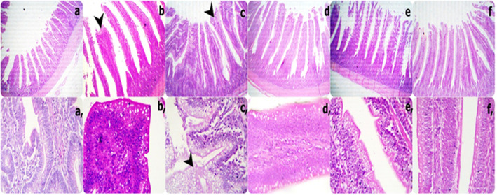
Representative photomicrograph of the low (40X) and high (400X) magnification H&E stained duodenums segment sections showing short and thick villi with limitation goblet cell metaplasia besides increasing inflammatory cells in the lamina propria in 0Vit.C+ 0Saff. Oil group; tall and thin villi with a few broad tips in a few villi without goblet cell metaplasia in 0Vit.C + 5 Saff. Oil group; marked brad tips and serrated surfaces with marked goblet cell metaplasia besides mild adhesions in 0Vit.C+ 10Saff. Oil group; more healthy which characterized by gradual increase tall and thin villi without goblet cells metaplasia in each 400Vit.C + 0Saff. Oil, 400Vit.C + 5Saff. Oil, and 400Vit.C + 10Saff. Oil groups respectively except increase intra-epithelium lymphocytic lick cells infiltrations in 400Vit.C+ 10Saff. Oil group. (a and a: 0Vit.C+ 0Saff. Oil group, b, and b: 0Vit.C + 5 Saff. Oil group, c, and c:0Vit.C+ 10Saff. Oil group: d and d: 400Vit.C + 0Saff. Oil, e and e: 400Vit.C + 5Saff. Oil, f and f: 400Vit.C+ 10Saff. Oil group)

