
Figure EV3. Examination of muscle fiber type‐specific dystrophin expression in treated mdx mice.
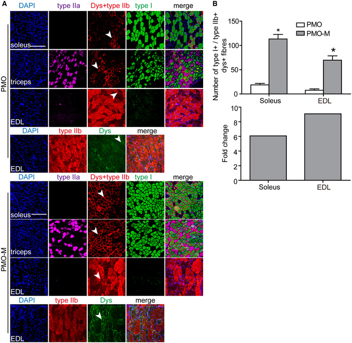
Adult mdx mice were intravenously injected with PMO at the dose of 12.5 mg/kg/week for 3 weeks mixed with MOTS‐c (500 μg), and tissues were harvested and examined two weeks after last injection.
- Immunohistochemistry for dystrophin expression in slow‐twitch (soleus), fast‐twitch (EDL), and mixed type (triceps) of muscle fibers of treated mdx mice (scale bar: 100 μm). Type I MHC was used to identify slow‐twitch muscle fibers, and type IIa and IIb MHC were used to characterize fast‐twitch muscle fibers. The arrowheads point to dystrophin‐positive fibers.
- Quantitative analysis of type I MHC‐ and dystrophin‐positive or type IIb MHC‐ and dystrophin‐positive fibers in treated mdx mice (n = 3; *P < 0.05, two‐tailed t‐test). Type I+ or type IIb+ means type I MHC‐positive or type IIb MHC‐positive fibers, respectively. Dys+ represents dystrophin‐positive fibers. Fold change refers to PMO‐M relative to PMO alone.
Data information: Data were presented as mean ± sem. Exact P values are specified in Appendix Table S1.