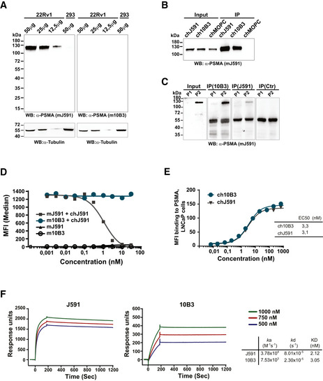
Figure EV1. In vitro characterization of the PSMA antibodies 10B3 vs J591.

-
AWestern blot analysis was performed using the indicated amounts of protein extracts prepared from 22Rv1high cells or from PSMA‐negative HEK‐293 cells. Blots were analyzed using anti‐PSMA (mJ591 or m10B3) and anti‐tubulin as a loading control.
-
B, CPSMA protein was immunoprecipitated from 22Rv1high cells (B) and lung SCC samples (C) using chimeric J591, 10B3, and MOPC‐21 (control) antibodies. The inputs (5%) and immunoprecipitates (30%) were analyzed by Western blot using murine J591 antibody.
-
DMouse Sp2/0 cells transfected with human PSMA were incubated with the indicated concentrations of primary murine mAbs (mJ591, m10B3), followed by a chimeric J591 antibody (chJ591, 10µg/ml) and finally with fluorescence labeled mouse anti‐human antibody for specific detection of chJ591 as indicated.
-
E, FChimeric versions of both mAbs were bound to LNCaP cells (E) and to a protein A coated sensor chip (F) and analyzed by flow cytometry and SPR, respectively, as described in the Materials and Methods section.
Source data are available online for this figure.
