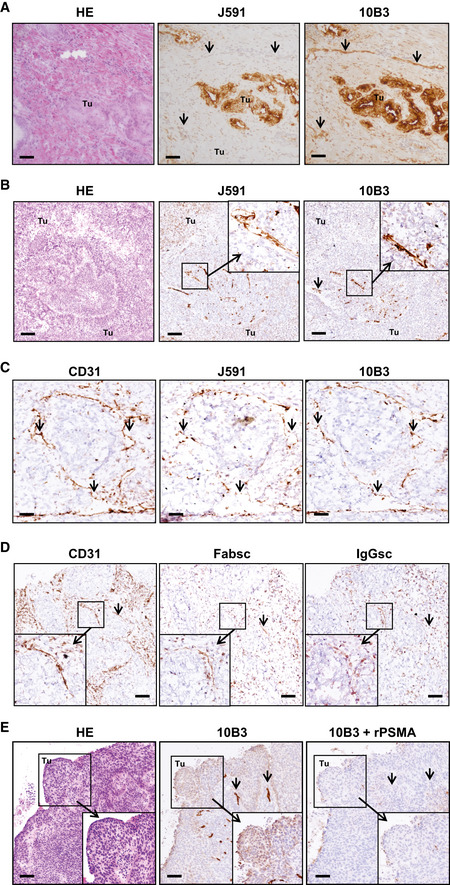
Figure EV2. Staining of exemplary prostate carcinoma and lung SCC samples.

-
ADirectly consecutive 3‐μm sections obtained from prostate carcinoma samples were stained with 10B3 and J591 mAbs. Arrows point to vessels. Tu: tumor. HE: Hematoxylin/Eosin staining. Scale 30 µm.
-
B–EDirectly consecutive 3‐μm sections (obtained from the same lung SCC sample for each antibody panel) were analyzed by immunohistochemistry as described in the Materials and Methods section. Note that in (C, D) a tumor sample with predominant vascular expression of PSMA (cancer cells PSMA‐very low intensity) was chosen to facilitate assessment of vascular staining. (B) Binding of murine 10B3 or J591 mAbs. Arrows point to vessels. Tu: tumor. HE: Hematoxylin/Eosin staining. Scale 30 µm. (C) Staining with 10B3, J591, or anti‐CD31 as marker for vessels. Arrows point to vessels. Scale 10 µm. (D) Binding of anti‐CD31 as well as biotinylated IgGsc and Fabsc molecules. Note that staining intensity with bsAbs is lower compared to CD31 due to the necessity to utilize a different detection protocol, and bsAb may additionally bind to tumor‐infiltrating T cells, resulting in differential staining patterns. Arrows point to vessels. Scale 20 µm. (E) Staining with 10B3 in the absence or presence of 50 µg recombinant PSMA protein. HE: Hematoxylin/Eosin staining. Scale 40 µm.
Source data are available online for this figure.
