
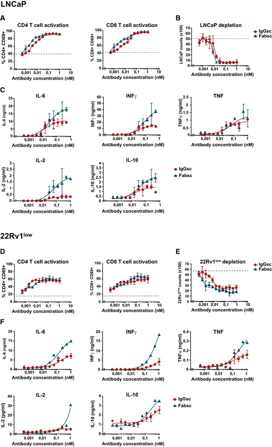
Figure 3. In vitro activity of the Fabsc and IgGsc molecules against tumor cells expressing different amounts of PSMA.
-
A–FBsAb were incubated at the indicated concentrations with PBMC of healthy donors together with LNCaP (A–C) or 22Rv1low cells (D–F) expressing high and low amounts of PSMA, respectively. After 3 days, CD4+ and CD8+ T‐cell activation (A, D), target cell depletion (B, E), and cytokine release (C, F) were determined by flow cytometry as described in the Materials and Methods section. Mean values and standard deviations of triplicate measurements are indicated. The dotted line represents the baseline T‐cell activation (A, D) or tumor cell counts (B, E) in the absence of bispecific antibodies.