
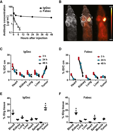
Figure 5. Half‐life and tumor localization of the Fabsc and the IgGsc molecules in immunodeficient mice.
-
A20 µg of the indicated bsAbs was injected i.v. into 57BL/6 mice, and serum concentrations were measured at the indicated time points after injection using the Promega assay described in the Materials and Methods section. Mean values and standard deviations obtained from groups of four animals per time point are indicated.
-
B64Cu labeled bsAbs were injected into SCID mice carrying established DU145 (PSMA‐negative, marked by “1”) and 22Rv1high (PSMA‐positive, marked by “2”) tumors on opposite flanks. MRT and PET scans were obtained after 48 h.
-
C–F64Cu labeled bsAbs were injected into NSG mice carrying 22Rv1high tumors (five animals per group), and uptake at the indicated locations was determined by PET quantification over time (C, D) and at termination of the experiment after 48 h (E, F) as described in the Materials and Methods section. Error bars represent standard deviation of the mean value.