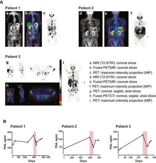
Figure EV5. PET scans and long‐term PSA levels of the three treated patients with castrate resistant, metastasized prostate carcinoma.

- Pre‐therapeutic PSMA expression of metastatic prostate cancer lesions was visualized by means of contrast enhanced whole body multiparametric PET/MRI (patient 1 and 2) or PET/CT (patient 3). Image data were acquired 60 min after i.v. injection of [18F]‐PSMA‐1007 (250–325 MBq), a labeled peptide tracer for PET imaging that specifically binds to PSMA. It is used according to §13.2B AMG (German drug law) for PSMA‐PET which has become standard clinical care in Germany. Representative images of the patients are shown: patient 1 presented with osseous and lymphonodal metastases with intense PSMA expression (a–c); patient 2 suffered from PSMA‐expressing peritoneal carcinomatosis, multiple PSMA‐expressing bone and lymph node metastases (d–f); in patient 3, osseous and lymphonodal metastases as well as local recurrence with intense PSMA expression are detected (g–i).
- Long‐term PSA values monitored prior, during (highlighted in light red), and after CC‐1 therapy. After documented failure of established treatment, patients were free of disease‐specific therapy for at least 4 weeks prior to application of CC‐1.
