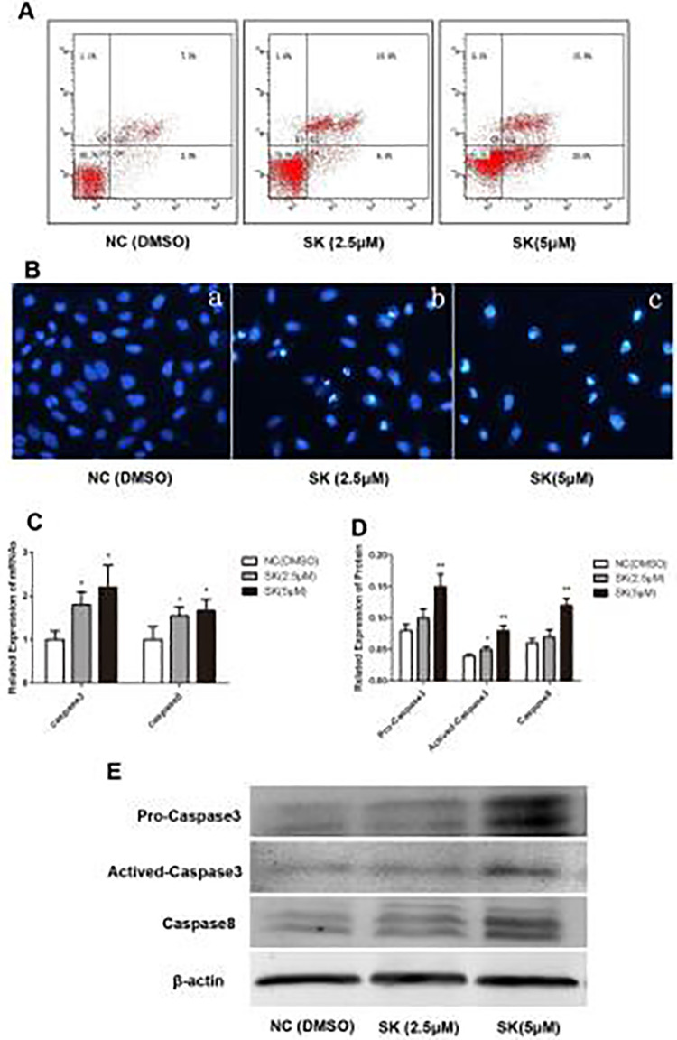
Figure 2.
Effect of Shikonin on cell apoptosis. (A) Apoptosis and necrosis of QBC939 were measured by flow cytometry with Annexin V/propidium iodide assay. (B) Morphological changes visualized under the fluorescence microscope with Hoechst 33342 staining (magnification 400×). (C) Changes of caspase-3 and caspase-8 mRNA expression were analyzed by real-time polymerase chain reaction (*P < 0.05, compared with the control). (D, E) Expression of pro-caspase-3, activated-caspase-3, and caspase-8 analyzed by Western Blot. (*P < 0.05, **P < 0.01, compared with the control).

