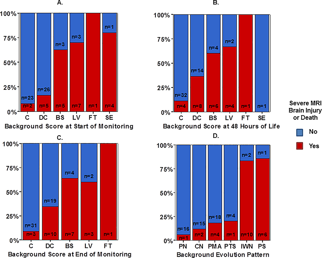
Figure 1:
Distribution of aEEG background pattern at the (A) start of monitoring, (B) 48 HOL, and (C) end of monitoring compared with overall aEEG background evolution classification (D), stratified by outcome group defined as severe MRI injury or death (red bar) or survived with normal/mild MRI (blue bar).

