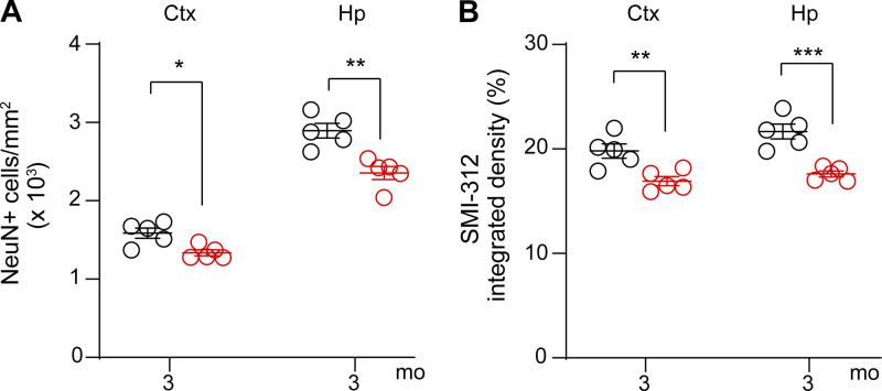
Figure S3.
Neuronal degeneration in 3-mo-old Lrp1lox/lox; Tie2-Cre mice after endothelial-specific Lrp1 deletion. (A and B) Quantification of NeuN+ neurons (A) and SMI312+ neurites (B) in the cortex (Ctx) and hippocampus (Hp) of 3-mo-old Lrp1lox/lox; Tie2-Cre and Lrp1lox/lox mice. Mean ± SEM, n = 5 mice/group. Significance by Student’s t test, *, P < 0.05; **, P < 0.01; ***, P < 0.001.

