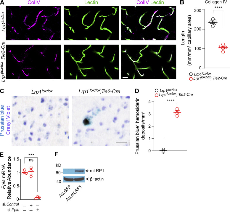
Figure S4.
Loss of collagen IV and hemosiderin deposits in Lrp1lox/lox; Tie2-Cre mice and additional characterization of reagents used in Fig. 4. (A and B) Immunostaining for collagen IV (CollV; magenta) and fluorescent staining for lectin+ endothelium (green) in the cortex of 2-mo-old Lrp1lox/lox;Tie2-Cre and Lrp1lox/lox control mice (A), and quantification of collagen IV length on brain capillary (<6 µm in diameter) lectin+ endothelial profiles in these mice (B). Scale bar = 25 µm. Mean ± SEM, n = 5 mice/group. (C and D) Prussian blue hemosiderin deposits in 2-mo-old Lrp1lox/lox;Tie2-Cre mouse and lack of Prussian blue deposits in Lrp1lox/lox control (C), and quantification of Prussian blue+ hemosiderin deposits in these mice (D). Scale bar = 20 µm. Mean ± SEM, n = 3 mice/group. (E) Inhibition of Ppia mRNA (encoding CypA) by siPpia, but not siControl, in brain endothelial cells isolated from 2-mo-old Lrp1lox/lox; Tie2-Cre mice. (F) Representative immunoblotting of mLRP1 in brain endothelial cells from 2-mo-old Lrp1lox/lox; Tie2-Cre mice after adenoviral-mediated reexpression with Ad.mLRP1 but not control Ad.GFP virus. Significance by Student’s t test; ns, not significant; ***, P < 0.001; ****, P < 0.0001.

