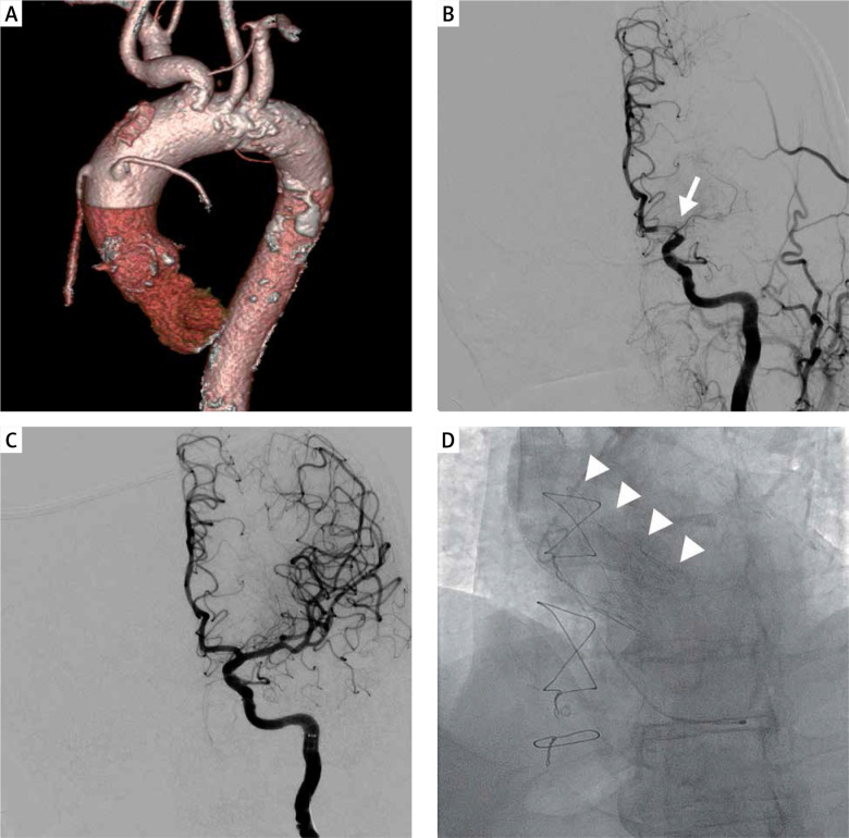
Figure 1.
A – Calcifications in sinotubular junction, aortic arch and descending aorta visible on pre-procedural computed tomography. Aortic valve calcification Agatston score was 2666. B – Cerebral angiography revealing occlusion of the left MCA at the proximal M1 segment (arrow). C – Follow-up angiography after mechanical thrombectomy confirming complete recanalization of left MCA. D – Final angiography showing correct position of the ACURATE neo L valve

