Figure 5.
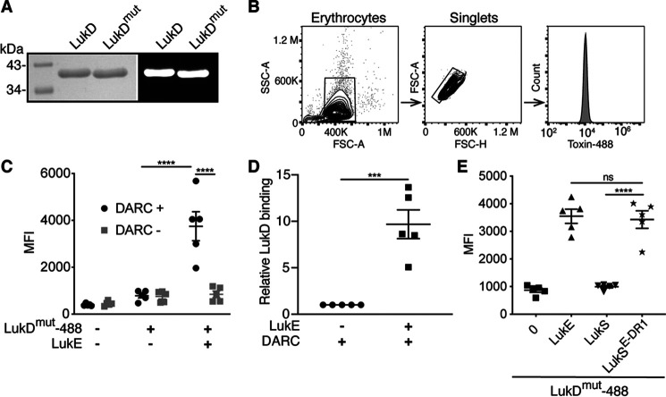
LukE promotes LukD binding to erythrocytes through DR1. A, left panel, Coomassie Blue–stained gel of DyLight 488–labeled toxins. Right panel, fluorescent image of gel. B, flow cytometry gating strategy of human erythrocytes used in C–E. C and D, binding of LukDmut-488 in the presence or absence of LukE with or without DARC. C, MFI. D, ratio of MFI relative to binding of LukDmut-488 alone after subtracting the background fluorescence of unstained cells (n = 5 donors). E, binding of LukDmut-488 in the presence of the indicated S subunits (n = 5 donors). LukDmut indicates LukDΔ130-4 (pore-formation incompetent), 488 indicates DyLight 488, and LukSE-DR1 indicates LukS-PV with the DR1 from LukE. The data are pooled from five (C and D) or two (E) independent experiments, and means ± S.E. are shown. ****, p < 0.0001; ***, p < 0.001; ns, not significant. C, two-way ANOVA with Sidak's correction. D, Student's t test. E, one-way ANOVA with Tukey's correction.

