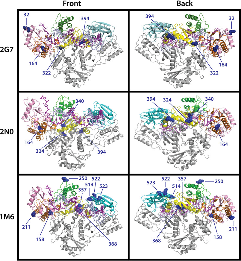
Figure 6.
Location of mutated residues in 2G7, 2N0, and 1M6 Vpx (−) RT variants. The crystal structure of heterodimeric HIV-1 RT bound to nucleic acid (PBD ID: 1RTD) was used to map the location of the equivalent mutated residues (blue spheres) in 2G7 (first row), 2N0 (second row), and 1M6 (third row) SIVmac239 Vpx (−) RT variants. Front and back views of the polymerase are displayed in the first and second column, respectively. The p66 fingers (pink), palm (orange), thumb (green), connection (yellow), and RNaseH (cyan) subdomains and p51 (gray) subunits are colored accordingly.

