Figure 5.
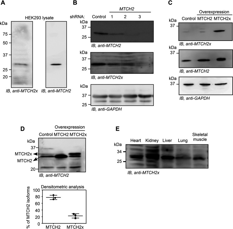
Detection of endogenous MTCH2x. A, Western blotting performed using an antibody (anti-MTCH2x) generated against the peptide encoded by the ISR1 (RCGAGTVTFL) in HEK293 cell lysate. B, Western blotting performed using the same antibody in HEK293 cells transfected with MTCH2-specific shRNAs. C, detection of exogenous overexpressed MTCH2x by anti-MTCH2x antibody in HEK293 cells. D, amount of MTCH2x relative to MTCH2 was estimated by performing a prolonged SDS-PAGE followed by Western blotting using anti-MTCH2 antibody that can potentially recognize all isoforms. Quantification is shown below (mean ± S.D., n = 3). Lysates from cells overexpressing MTCH2 or MTCH2x (without any tag) were used to distinguish endogenous MTCH2 and MTCH2x. E, detection of endogenous MTCH2x in mitochondrial extracts from multiple mouse organs.

