Figure 7.

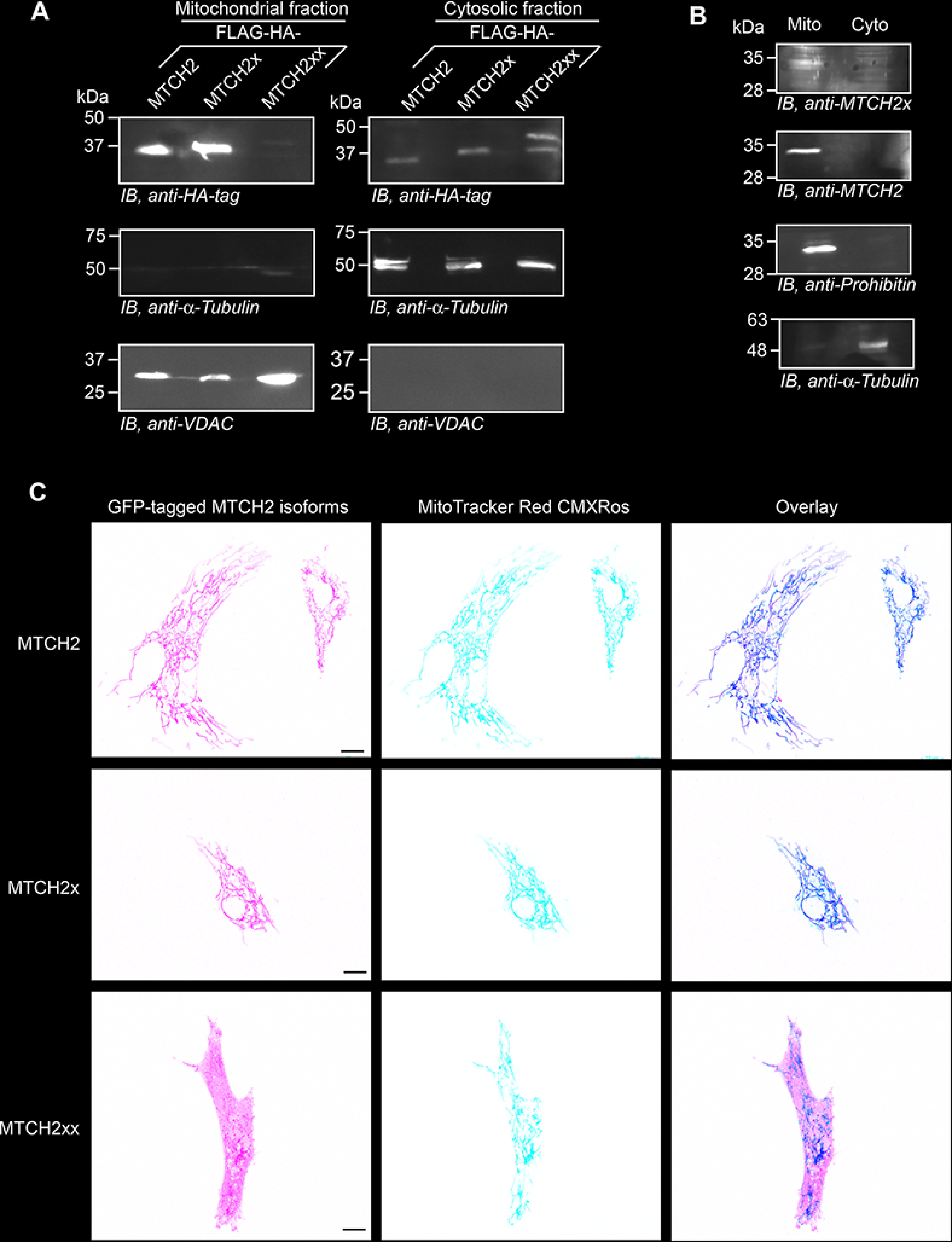
Differential localization of MTCH2 isoforms. A, plasmids expressing FLAG-HA–tagged isoforms of MTCH2 were transfected in HEK293 cells. Mitochondrial and cytoplasmic fractions from these cells were used for Western blotting. VDAC and α-tubulin were used as markers for mitochondria and cytoplasm, respectively. B, mitochondrial localization of endogenous MTCH2 and MTCH2x in HEK293 cells. Prohibitin was used as a marker for mitochondria. C, confocal fluorescence microscopy images showing cellular localization of GFP-tagged MTCH2 isoforms in primary bovine aortic endothelial cells. MitoTracker Red CMXRos was used to stain mitochondria. Percentage of GFP-positive cells in case of GFP-MTCH2xx was less compared with GFP-MTCH2 and GFP-MTCH2x. One such cell is shown. Scale bar, 10 μm.
