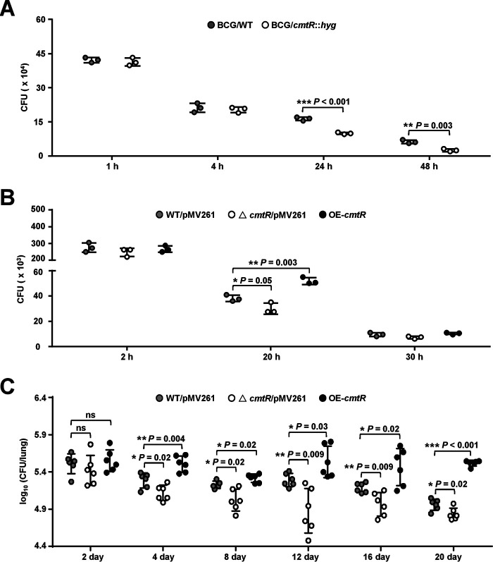
Figure 7.
Assays for studying the effect of CmtR on intracellular survival of mycobacteria in macrophages and in mice. Cells were infected with mycobacterial strains at a multiplicity of infection of 10 and washed three times at 4 h post infection to remove extracellular bacteria. Thereafter, the cells were re-incubated in a medium supplemented with penicillin/streptomycin and lysed using 0.025% SDS at the indicated post-infection time points. Serial dilutions of the supernatant were then plated on 7H10 agar supplemented with 10% oleic acid–albumin–dextrose–catalase, and the number of cfu (CFU) was counted 15–21 days later. A, RAW264.7 cells were infected with BCG/WT and BCG/cmtR::hyg strains separately. B, BMDMs were infected with WT/pMV261 (BCG/pMV261), △cmtR/pMV261 (BCG cmtR::hyg/pMV261), and OE-cmtR (BCG/pMV261-cmtR) strains, respectively. C, female SPF C57BL/6 mice (n = 6 mice per group) were infected intratracheally with 1 × 10⁶ of WT/pMV261, △cmtR/pMV261, and OE-cmtR strains for 0–20 days, and the bacterial loads in lung tissue homogenates of mice were determined. Error bars represent the S.D. from three biological experiments. The P-values of the data were calculated by unpaired two-tailed Student's t test using GraphPad Prism 7. Asterisks represent significant difference (*, P < 0.05; **, P < 0.01; ***, P < 0.001; ns, not significant; two-tailed Student's t test) between two groups.

