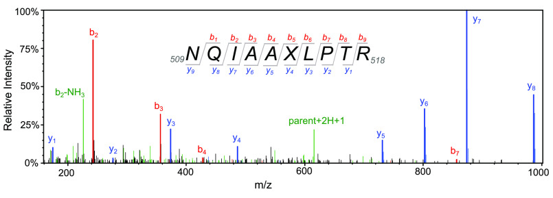
Figure 6.
MS/MS spectrum of tryptic peptide containing 7-HCou. Tryptic peptides from gel slices containing T7 DNA polymerase E514Cou were resolved by reverse-phase HPLC and analyzed by MS/MS to confirm successful incorporation of 7-HCou. A representative MS/MS spectrum after collision-induced dissociation of the tryptic peptide corresponding to amino acids 509–518 is shown. Collision-induced dissociation most often produces b ions or y ions, with intact N or C termini, respectively. The subscript number following the letter indicates the number of amino acid residues in the ionized peptide fragment. Peaks identified as b or y ions for the parent peptide 509NQIAAXLPTR518 are shown in red and blue, respectively. The parent peptide (+2H +1) and the b2 ion (−NH3) are shown in green. X denotes the position of 7-HCou in the peptide, with a mass shift of +118 Da relative to glutamic acid in the WT enzyme.

