Figure 3.
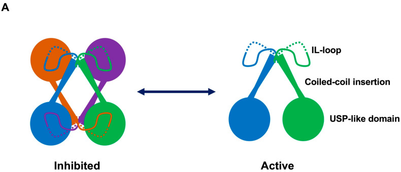
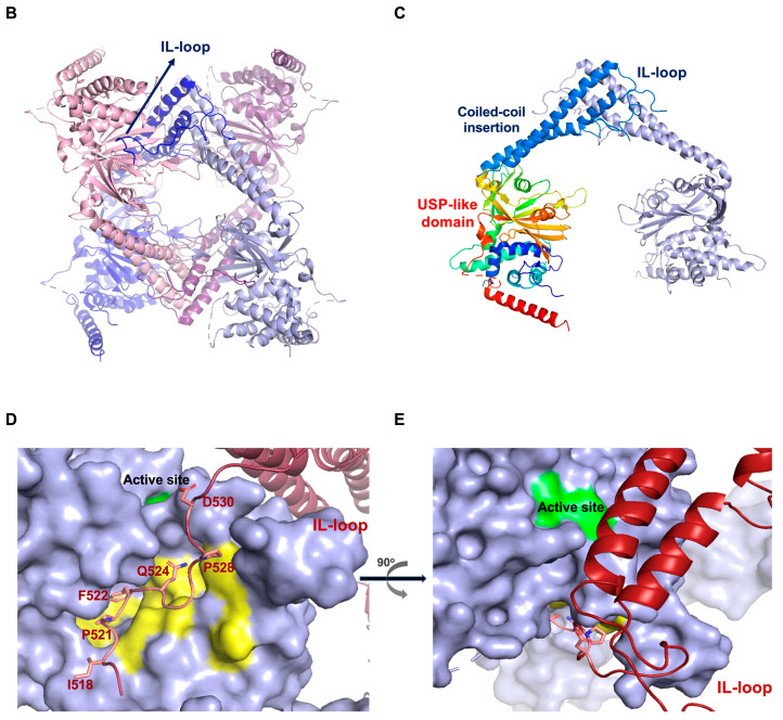
The structure of USP25. (A) Schematic cartoon representation of the USP25 tetramer and dimer assemblies. Each monomer is colored. (B) Crystal structure of the USP25 tetramer (PDB: 5O71 and 6HEL). Each dimer is colored blue or light pink. (C) Crystal structure of the USP25 dimer. IL-loop, coiled-coil insertion and ubiquitin-specific protease (USP)-like domain are marked. (D) Binding interface of the IL-loop with the ubiquitin-binding site of USP25. IL-loop (red), active site (green), binding surface (yellow), the interface contacts of IL-loop are labelled. (E) View of D rotated 90°.


