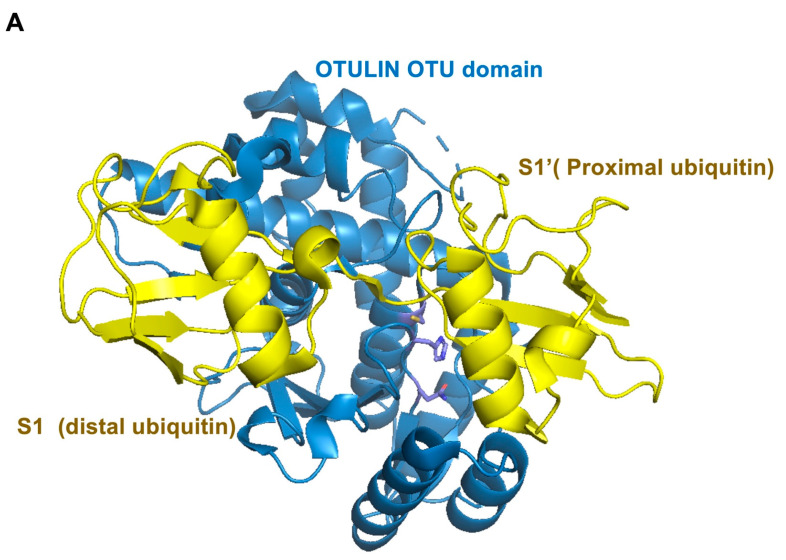
Figure 5.
The structure of OTULIN. (A) Structure of OTULIN OTU domain in complex with Met1-di-ubiquitin. OTULIN (blue), ubiquitin (yellow). (B) Rearrangement of the active site residues in the absence (white blue) of bound Met1-di-ubiquitin. (C) Rearrangement of active site residues in the presence (blue) of bound Met1-di-ubiquitin. (D) Superposition of (B,C). (PDB: 3ZNZ and 3ZNV).


