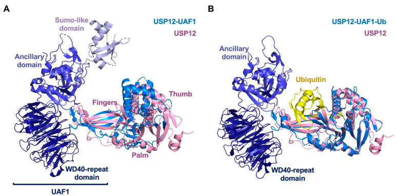
Figure 7.
Structure of USP12-UAF1-WDR20. (A) Structure of with USP12-UAF1. (B) Structure of with USP12-UAF1(9-580)-Ub. (C) Active site of USP12. (D) Differences of Fingers domain in the absence and presence of UAF1. (E) Structure of USP12-UAF1(9-580)-WDR20. Free USP12 (pink), UAF1(deep blue), WDR20 (green), ubiquitin (yellow). (PDB: 5K16, 518W, 5K1B, and 5K1C).


