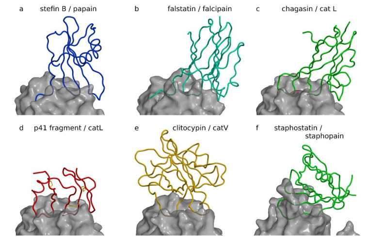
Figure 1.
Inhibitors of papain-like and related proteases. Complexes are shown with the same view across the active site cleft and the same scale after superimposition of proteases to cathepsin L in the p41 fragment complex. Figure was prepared using MAIN [35] and rendered with Raster3d [36]. (a) Stefin B papain complex ([21], PDB code 1STF). The stefin B chain is shown as a blue coil on the semitransparent background of the white surface of papain. (b) Inhibitor of cysteine protease (ICP) (falstatin) falcipain complex ([37], PDB code 3PNR). ICP, also known as falstatin from Plasmodium berghei, is shown as a cyan coil on the semitransparent background of the white surface of falcipain-2. (c) Chagasin cathepsin L complex ([38], PDB code 2NQD). The chagasin chain is shown as a green coil on the semitransparent background of the white surface of cathepsin L. (d) p41 fragment cathepsin L complex ([39], PDB code 1ICF). p41 fragment chain shown as a red coil on the semitransparent background of the white surface of cathepsin L. The three disulfide bonds of the p41 fragment are shown as yellow sticks. (e) Clitocypin cathepsin V complex ([40], PDB code 3H6S). The clitocypin chain is shown as a yellow coil on the semitransparent background of the white surface of cathepsin V. (f) Staphostatin staphopain complex ([41], PDB code 1PXV). The staphostatin chain is shown as a green coil on the semitransparent background of the white surface of staphopain.

