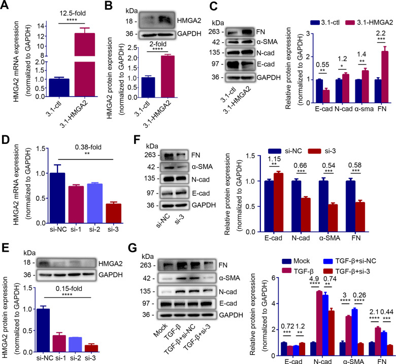
Figure 2.
HMGA2 contributes to epithelial-mesenchymal transition (EMT) of endometrial epithelial cells. (A) HMGA2 mRNA level (n = 5) and (B) Left: western blot analysis of HMGA2 protein level in Ishikawa (IK) cells transfected with HMGA2 high expression plasmid or control plasmid for 24 h. (C) Left: western blot analysis of E-cadherin (E-cad), N-cadherin (N-cad), α-smooth muscle actin (α-SMA) and fibronectin (FN) proteins levels in IK cells transfected with HMGA2 high expression plasmid or control plasmid for 48 h. (D) HMGA2 mRNA expression (n = 3), and (E) Top: western blot analysis of HMGA2 protein level in IK cells transfected with different small interfering RNAs of HMGA2 (si-1, si-2, si-3) or negative control (si-NC) for 24 h. (F) Left: western blot analysis of E-cad, N-cad, α-SMA and FN proteins levels in IK cells transfected with si-HMGA2 or si-NC for 48 h. (G) Left: western blot analysis of E-cad, N-cad, α-SMA and FN proteins levels in TGF-β-pretreated IK cells transfected with si-HMGA2 (TGF-β + si-3) or si-NC (TGF-β + si-NC) for 48 h. (C, F, G) Right and (E) Bottom: quantification of corresponding proteins (n = 3) determined by image J software. The data are shown as the mean ± SD. *P < 0.05; **P < 0.01; ***P < 0.001; ****P < 0.0001. Student’s t-tests were performed for all panels. The number above the asterisk in each panel is the fold difference.

