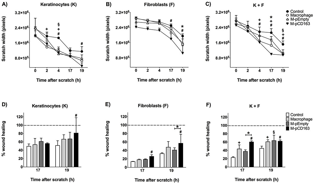
Figure 6. Wound healing assay and migration temporal course of keratinocytes and/or fibroblasts cultures in the presence of CD163-overexpressing THP-1 macrophages.
Time course (time-response curves) of the migration healing progression of keratinocytes (A), fibroblasts (B), keratinocytes + fibroblasts − K +F (C) alone (Control) or in the presence of LPS-stimulated non-transfected macrophages (macrophage), pEmpty-transfected macrophages (M-pEmpty) or pCD163-transfected macrophages (M-pCD163) cultures at 0, 2, 4, 17 and 19 hours after the scratch. Comparison of wound healing (%) between keratinocytes (D), fibroblasts (E), or K + F (F) alone (control) or in the presence of LPS-stimulated non-transfected macrophages (macrophage), M-pEmpty or M-pCD163 at 17 and 19 hours after the scratch. Macrophages were previously stimulated with LPS. The closure of the scratch gap (scratch width) was measured in pixels. N= 3 per group (A-F). +§# p<0.05 all groups vs. control group; *p<0.05 M-pEmpty vs. M-pCD163 group, by Two-way ANOVA and Bonferroni post-test. 2-column fitting image

