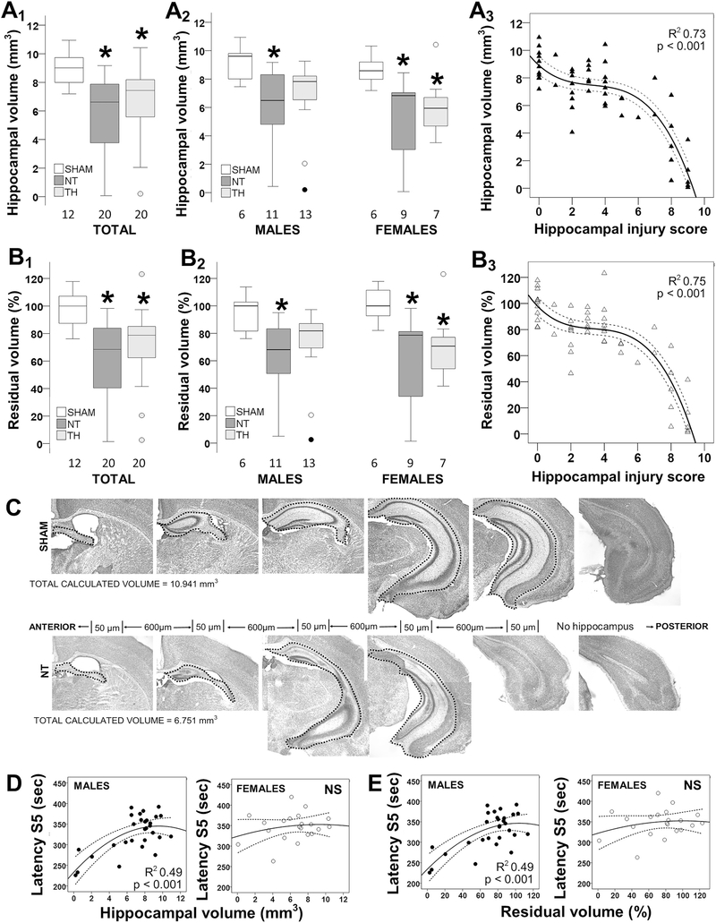
Fig. 1.
Correlation between hippocampal atrophy after neonatal hypoxia-ischemia (HI) and seizure susceptibility. Hippocampal volume (A) and residual volume (B) are decreased in mice exposed to HI and normothermia (NT) and this decrease is attenuated by TH (A1, B1), which is sexually dimorphic (A2, B2). Results are shown as box and whisker plots, where the box is limited by the 25th and 75th percentiles (interquartile range, IQR) and the solid line represents the median. The whiskers are limited by the last data point within 1.5 times the IQR from the median, with outliers included. Kruskal–Wallis ANOVA with Dunn-Bonferoni post-hoc testing for pair analysis was applied. *, p < 0.05. The relationship between hippocampal volumes and histological injury scores is not linear (A3, B3). Representative photomicrographs are shown demonstrating hippocampal volume measurements (C). Hippocampal volumes and residual volumes correlated directly with latency to S5 in male mice (D, E). The continuous lines represent the fitted line derived from a non-linear regression and the discontinuous lines represent the 95% confidence boundaries.

