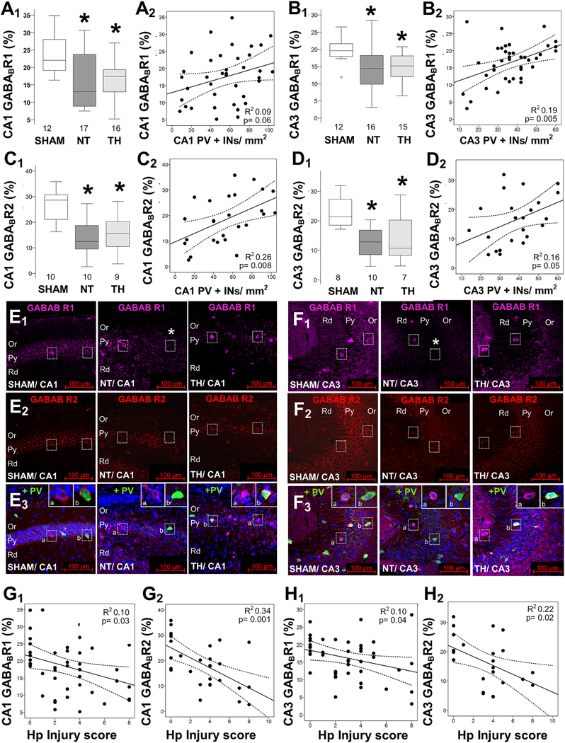
Fig. 3.
Decreased GABAB receptor subunits in the hippocampus after neonatal HI. HI decreases GABAB R1 subunit (GABABR1; A, B) and GABAB R2 subunit (GABABR2; C, D) percent area of expression within the PCL of the CA1 (A1, C1) and CA3 (B1, D1). No sexual dimorphism was identified (data not shown). Boxes are limited by the 25th and 75th percentiles (interquartile range, IQR) and whiskers are limited by the last data point within 1.5 times the IQR from the median (continuous line inside the box), with outliers included. Kruskal–Wallis ANOVA with Dunn-Bonferoni post-hoc testing for pair analysis was applied. *, p < 0.05. The correlations between the number of PV+INs in hippocampal CA1 (A2, C2) and CA3 (B2, D2) with GABABR1 (A2, B2) and GABABR2 (C2, D2) are shown. Representative images from GABABR1 (Alexa 647, magenta) and GABABR2 (Alexa 568, red) combined with PV (Alexa 488, green) and nuclear stain with DAPI (blue) in merged image, were captured at 20×/0.8 objective to show the CA1 (E) and CA3 (F) hippocampal pyramidal (Py), oriens (Or), and radiatum (Rd) layers. GABABR1 and GABABR2 levels correlate with hippocampal injury scores in CA1 (G1,2) and CA3 (H1,2). The continuous lines represent the fitted line derived from a linear regression and the discontinuous lines represent the 95% confidence intervals. Sex-stratified and non-stratified correlations with fluorothyl susceptibility were non-significant (data not shown).

