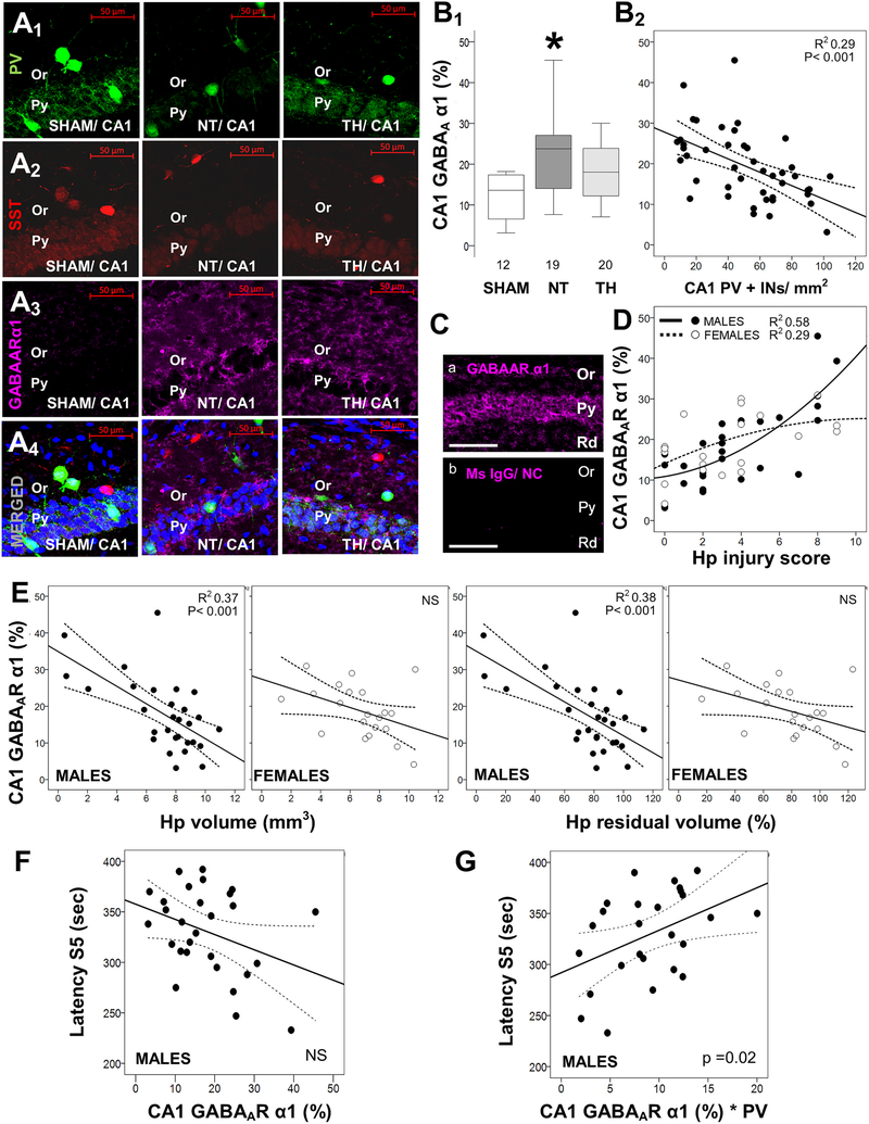
Fig. 4.
Increased hippocampal levels of GABAA receptor α1 subunit correlate with flurothyl seizure susceptibility. Shown are representative confocal images (20×/0.8 objective) of hippocampal pyramidal (Py) and oriens (Or) layers for PV (Alexa 488, green; A1), somatostatin (SST, Alexa 568, red; A2), GABAA receptor α1 subunit (GABAARα1, Alexa 647, magenta; A3), and merged (A4) with nuclear DAPI (blue) in sham, normothermia (NT), and therapeutic hypothermia (TH) mice. Box and whisker plots demonstrate the percent expression of GABAARα1 in each group (B1) and the correlations with the number of PV+ interneurons (INs) in the CA1 region (B2). In some instances, GABAARα1 immunoreactivity was extreme despite blocking methods (Ca) and absent of non-specific staining in negative controls (Cb). Correlations between GABAARα1 and hippocampal (Hp) injury scores (D), Hp volume measurements (E), and latency to stage 5 (S5) seizures with flurothyl (F) are shown. Adjustment of the number of PV+INs by their relative expression of GABAARα1 affects the correlation with latency to S5 seizures with flurothyl (G), now becoming linear. Boxes are limited by the 25th and 75th percentiles (interquartile range, IQR) and whiskers are limited by the last data point within 1.5 times the IQR from the median (continuous line inside the box), with outliers included. Kruskal–Wallis ANOVA with Dunn-Bonferoni post-hoc testing for pair analysis was applied. *, p < 0.05. In correlation charts, the continuous lines represent the fitted line derived from regression modeling and the discontinuous lines represent the 95% confidence boundries.

